Nursigo · Anatomy essentials
See the anatomy.
Make the connection.
Turn structures into understanding with focused organ exploration, short study notes and free knowledge checks. Start with an illustration, then open 3D when you want a closer look.
Renal · Visual study
Understand the kidney
Explore the cortex and medulla, then connect filtration to urine formation.
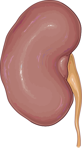
Optional 3D download: 0.4 MB. Study notes and questions work with the illustration too.
Right and left always refer to the person, not the viewer.
Choose a structure
Read its role below. Open 3D to isolate it in the model.
Start with the whole organ
Explore the cortex and medulla, then connect filtration to urine formation.
Follow urine leaving the kidney
- 1. Papilla
- 2. Minor calyx
- 3. Major calyx
- 4. Renal pelvis
- 5. Ureter
- 6. Bladder
Exam connection
Keep the two pathways separate: blood reaches the kidney through arteries; urine leaves through a ureter. The nephron is the functional unit, not the whole renal pyramid.
Learning sources & visual credits
Illustration: Servier Medical Art, unchanged. 3D: Human Reference Atlas, Visible Human Female, by Kristen Browne and Heidi Schlehlein; geometry optimized for the web. Both under CC BY 4.0. Full asset credits and changes.
Nursigo-authored foundation questions. These supplement your exam-specific practice; they are not official exam items.
Put the foundations into practice
Continue with questions for your nursing licensure or specialty exam.